Nonmalignant Dermatologic Diseases of the Male Genitalia: Introduction, Definition of Terms, and Anatomy ... - Medscape Reference
Balanitis xerotica obliterans
Balanitis xerotica obliterans (BXO), also known as lichen sclerosus, is an inflammatory disorder that usually affects the anogenital skin. Lichen sclerosus is a benign disorder of unknown etiology and is far more prevalent in women than men. In men, it is commonly referred to as BXO, as it usually involves the glans penis and the foreskin. Uncircumcised males are usually affected, and BXO is an established cause of phimosis and meatal stenosis in this population. Among those with BXO, complications often arise as a consequence of the sclerosis and scarring typical of this disorder.
Pathophysiology
The exact pathogenesis of BXO is unknown. Both an infectious and an autoimmune etiology have been proposed. An association with Borrelia burgdorferi has been reported in some series; however, this has not been substantiated in other reports. Although some patients with BXO are more likely to have an accompanying autoimmune disorder, pathogenic antibodies have not been consistently demonstrated in the affected skin or sera of those with this condition. BXO/lichen sclerosus may be a superficial variant of morphea (localized scleroderma) because they share similar histopathologic features, and the two conditions may coexist in the same patient.
Clinical presentation/diagnosis
BXO occurs in a wide age range, including young prepubescent boys and older men. Males with BXO typically present with phimosis and/or recurrent balanitis. In addition, pain during urination or erection may also be a presenting symptom. BXO usually involves the glans, prepuce, urethral meatus, and, occasionally, the shaft. [64, 65] The clinical presentation is that of well-defined, atrophic pale-gray to ivory-white macules or patches that resemble cigarette paper. Punctate areas of hemorrhage, fissures, and erosions are commonly apparent because of the pruritic nature of the lesions. Focal areas of hyperkeratosis may also develop.
The diagnosis can be established clinically; however, biopsy is often necessary for confirmation. The characteristic histopathologic findings include epidermal orthokeratosis and atrophy with focal vacuolar change of the basal layer and hyalinization (sclerosis) of the papillary and superficial reticular dermis. [66, 67, 68] A superficial perivascular lymphocytic inflammatory infiltrate is frequently present.
Differential diagnoses
The differential diagnoses include vitiligo (see Vitiligo), scar, and atrophic lichen planus. Symptoms are usually absent in patients with vitiligo. Patients with scars may have a history of prior trauma or inflammation. Patients with lichen planus frequently have characteristic lesions in other sites. Biopsy is often necessary to distinguish these disorders, as well as to exclude the possibility of early squamous cell carcinoma. [65]
Treatment and outcome
For early-stage disease, topical therapies may be effective. However, late-stage disease with extensive sclerosis is unlikely to respond well to medical therapy, and surgical intervention is often required. Circumcision is the therapy of choice for lesions that involve the foreskin. Topical agents, such as testosterone creams, petrolatum, and antifungals, are generally ineffective. Although high-dose topical corticosteroids have been used with success in women (in some cases even reversing the course of disease), they have not been studied in detail in men. Based on limited studies in men, topical corticosteroids may be effective, particularly in early disease. [67] However, long-term corticosteroid treatment may cause epidermal atrophy or lead to reactivation of latent infections such as HPV.
Other treatment modalities involving surgery, laser therapy, and retinoids produce limited success. [69, 70, 71] Topical tacrolimus is often effective. [72] If urethral involvement and stenosis are present, meatotomy or dilation may be necessary.
The outcome of BXO is variable and depends on the degree of sclerosis and scarring. Patients may require surgical intervention for structural or functional impairment. Meatal stenosis is a complication of urethral meatal involvement that may require surgical management.
BXO is associated with penile squamous cell carcinoma; however, the actual incidence of squamous cell carcinoma in patients with BXO is unclear. [73] One study showed that squamous cell carcinoma developed in 5.8% of patients with BXO. [74] Some studies have suggested that HPV may be a cofactor in the development of penile squamous cell carcinoma in patients with BXO. [75] However, other studies have shown non-HPV–associated penile squamous cell carcinoma arising in patients with BXO. In females, lichen sclerosus is also associated with vulvar squamous cell carcinoma in a minority of cases.
Lichen planus
Lichen planus is a pruritic papulosquamous skin disorder of unclear etiology. The skin, mucosal surfaces, and/or nails may be affected. Patients may have genital involvement along with widespread cutaneous disease, or the disease may be restricted to the genital and/or mucosal regions.
Pathophysiology
Although several hypotheses have been proposed, the specific etiology of lichen planus is unknown. Cell-mediated immune response and alteration of epidermal antigenicity appear to be important in the pathogenesis. Genetic factors play a role, as lichen planus is significantly associated with HLA-DR1 and HLA-DQ1. In recent years, an epidemiological association has been established between hepatitis C viral infection and lichen planus.
Clinical presentation/diagnosis
Usually, the onset of genital lichen planus occurs in the fourth to sixth decade of life. A possible delayed hypersensitivity reaction to an unknown epidermal antigen is the current theory. The cellular immune response to basal epithelial cells causes initial destruction of the basal cell layer by T cells. Familial cases and cases associated with liver cell abnormalities, including chronic active hepatitis and primary biliary cirrhosis, have been reported. HLA-DR1 is associated with lichen planus.
The classic presentation is that of widespread discrete, flat-topped, polygonal papules that have a predilection for the flexural surfaces. The individual papules are violaceous in color and often have overlying white lines known as Wickham striae. (See the images below.) The lesions tend to be extremely pruritic, and, as they heal, considerable dyspigmentation is notable at sites of prior involvement. Nails and the oral mucosa are frequently involved. Genital involvement is common in males, affecting up to 25% of patients with widespread disease.
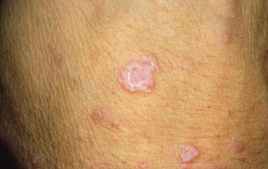 Lichen planus (Image courtesy of Hon Pak, MD)
 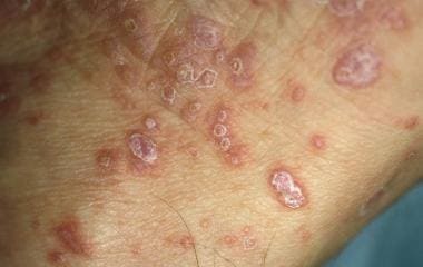 Lichen planus (Image courtesy of Hon Pak, MD)
 Occasionally, penile involvement may occur in the absence of other cutaneous lesions. In the genital region, the lesions often appear annular, eroded, or ulcerated. Patients may present with itching, pain, or dysuria. In the absence of characteristic cutaneous lesions, biopsy is necessary to establish the diagnosis. The histopathology shows epidermal hyperkeratosis, acanthosis, wedge-shaped hypergranulosis, and interface alteration of the basal keratinocytes. The dermis displays a superficial, dense bandlike, predominantly lymphocytic inflammatory infiltrate. Apoptotic keratinocytes, known as Civatte or cytoid bodies, are typically present.
Differential diagnoses
Complete evaluation of the oral mucosa, skin, and nails is often helpful in establishing the diagnosis of lichen planus. The differential diagnoses include psoriasis, candidiasis, fixed drug eruption, early erythema multiforme, herpesvirus infection, syphilis, Zoon balanitis, BXO, erythroplasia of Queyrat, and squamous cell carcinoma. Most of these conditions can be differentiated on clinical grounds; however, microbiologic culture, skin biopsy, and serologies may be necessary for diagnosis in some cases.
Treatment and outcome
The standard therapy for genital lichen planus is topical corticosteroids. Symptoms and clinical response generally improve within several weeks. When severe, systemic corticosteroids (ie, prednisone) in doses ranging from 30-40 mg daily, tapered over 6-8 weeks, can be helpful. In steroid-resistant cases, therapy with systemic retinoids or topical immune modulators such as tacrolimus, pimecrolimus, [76] or cyclosporin may be helpful.
In most cases, lichen planus resolves spontaneously within 1-2 years. Recurrence and remissions separated by years are not uncommon. Scarring in the form of dyschromia—both hyperpigmentation and hypopigmentation—is a characteristic sequela of this disease. Phimosis due to lichen planus has been reported and may require circumcision if severe or unresponsive to medical therapy. Penile squamous cell carcinoma arising in lesions of lichen planus has been very rarely reported.
Fixed drug eruption
Fixed drug eruptions are erythematous lesions that develop within hours after a drug is taken and recur at the same site upon subsequent exposure to the same drug (see Drug Eruptions). [77] The lesions may be solitary or multiple and show a predilection for the lips, extremities, and genitalia. A large number of drugs have been reported to elicit fixed drug eruptions. The most common drugs implicated include sulfonamides, tetracyclines, trimethoprim-sulfamethoxazole (TMP-SMZ), ibuprofen, phenolphthalein, barbiturates, acetyl-salicylic acid, and quinine. An increased susceptibility is associated with HLA-B22. [78]
Pathophysiology
The specific pathogenic mechanism underlying the recurrent lesions in fixed drug eruptions is not fully understood. Current concepts of pathogenesis have focused on cell-mediated responses that involve effector CD8+ T cells. In addition, the recurrence at a fixed site appears to be related to memory T cells at the lesional site. Recent evidence has shown that apoptosis of keratinocytes by CD8+ T cells may play an important role in the pathogenesis of fixed drug eruptions.
Clinical presentation/diagnosis
Fixed drug eruptions present as erythematous to violaceous plaques that develop 30 minutes to several hours after a drug is taken. Some lesions show vesiculation. Rare cases of widespread bullous fixed drug eruption, clinically resembling toxic epidermal necrolysis (TEN), have been reported. Lesions typically involve the lips, hands, feet, or genitalia. The glans is the typical site involved in penile lesions. Pruritus and burning may be associated symptoms. The lesions resolve in days and leave a hyperpigmented brown macule that eventually fades. Re-exposure to the offending drug elicits lesions at the same site. Additional sites may become involved over time. The clinical history and characteristic presentation are often sufficient for diagnosis; however, diagnosis is often confirmed with biopsy.
The histopathologic features of fixed drug eruption show interface alteration of the basal epidermis with focal necrotic keratinocytes, a superficial dermal mixed inflammatory infiltrate with scattered eosinophils, and pigment incontinence in the papillary dermis.
Differential diagnoses
The clinical differential diagnoses of penile fixed drug eruption include erythema multiforme, lichen planus, and immunobullous diseases. In the widespread bullous variant of fixed drug eruption, TEN is in the differential diagnoses.
Treatment and outcome
Cessation of the associated drug induces resolution of lesions. Topical corticosteroids can be used to treat symptomatic lesions.
Recurrence is common if the patient is re-exposed to the offending drug. Generally, the outcome is excellent with no significant long-term complications. Postinflammatory hyperpigmentation may present a cosmetic concern in some patients.
Psoriasis
Psoriasis is a chronic skin disorder that affects approximately 2% of the population. The most common form of psoriasis is plaque-type psoriasis, which is characterized by the presence of well-demarcated, silvery-scaled, erythematous plaques. A significant percentage (5-42%) of patients with psoriasis may also have arthritis, which may be debilitating if it is severe.
Pathophysiology
The specific pathogenic mechanisms underlying psoriasis are not fully understood, but several factors, including genetic and environmental factors, play a role. A polygenic inheritance pattern has been proposed, as a positive family history is detected in up to one third of patients. HLA antigens, specifically HLA-Cw6 and HLA-DR7, confer a greater relative risk of developing psoriasis. Infections (streptococcal pharyngitis in particular), psychogenic stress, medications, and cutaneous injury have all been identified as triggers of psoriasis, particularly in genetically predisposed individuals.
Experimental evidence suggests that psoriasis is a T-lymphocyte–mediated skin disease. In addition, several reports have demonstrated an altered expression of cytokines, particularly interleukin (IL)–10, tumor necrosis factor (TNF)–a, and IL-8, in psoriatic skin lesions. The imbalance of cytokines likely plays a role in the acceleration of the keratinocyte turnover time in the epidermis of psoriatic lesions. Polymorphonuclear cells are also thought to be involved in the pathogenesis of psoriasis, although their exact role is unclear.
Clinical presentation/diagnosis
Psoriasis may present in individuals of any age, but most patients present before the fifth decade of life. Several variants of psoriasis exist, and any area of the skin may be affected. The most common form of psoriasis is chronic plaque-type psoriasis. Sites of predilection include the knees, elbows, and scalp; however, the trunk, nails, intergluteal fold, and genitals may also be involved. The classic psoriatic lesion is that of a sharply marginated, erythematous plaque with silvery scale.
In the pustular form of psoriasis, lesions are characterized by erythematous macules or patches with overlying pinpoint sterile pustules. This and other forms of psoriasis (erythrodermic, guttate, inverse) do not exclusively affect the genitals. Nail involvement may appear as nail plate thickening, yellow-brown discoloration ("oil staining"), and/or separation of the nail plate from the nail bed (onycholysis). In a significant number of males with plaque-type psoriasis, genital involvement may occur, but the genital region is rarely the exclusive site of involvement. Any portion of the scrotum or penis may be affected. In the genital region, rather than appearing as distinctly elevated, silvery-scaled plaques, the lesions often have less scale and are barely raised (or nonraised) erythematous lesions.
Pruritus is frequently present but is typically not severe. Genital psoriasis can usually be diagnosed on clinical grounds. Because most patients also have nongenital involvement, thorough skin examination, particularly of the scalp, elbows, knees, nails, umbilicus, and intergluteal fold, is necessary. If characteristic psoriatic lesions are lacking, a skin biopsy may be necessary for confirming the diagnosis.
The histopathologic features of a well-developed psoriasis lesion show epidermal hyperparakeratosis, hypogranulosis, acanthosis, and regular psoriasiform hyperplasia with thinning of the suprapapillary plate. Intracorneal and intraepidermal microabscesses are focally present. The dermis displays dilated vascular channels in the papillary dermis and a superficial, mild-to-moderate, mixed inflammatory infiltrate. Lesions of pustular psoriasis typically exhibit more extensive intraepidermal microabscesses and less pronounced acanthosis and hyperplasia of the epidermis.
Differential diagnoses
The differential diagnoses of psoriasis involving the male genitalia include candida, tinea, seborrheic dermatitis, lichen planus, syphilis, Zoon balanitis, and early pemphigus. If the patient has systemic symptoms, reactive arthritis should be considered. If a solitary lesion is present, skin biopsy may be necessary to exclude the possibility of erythroplasia of Queyrat, Bowen disease, or squamous cell carcinoma.
Treatment and outcome
Low- to mid-potency topical corticosteroids are the mainstay of therapy for genital psoriasis. With long-term topical corticosteroid application, atrophy can occur and may limit its use in this particular body region. Topical therapy with calcipotriol (see calcipotriene), a vitamin D analogue, or calcineurin inhibitors such as tacrolimus or pimecrolimus are also potential options. These latter therapies can be used alone or in combination with topical corticosteroids to minimize adverse effects. Other topically applied medications include the keratolytic agent, salicylic acid, and the retinoid tazarotene. The irritant effect of the latter often limits its use in the genital area.
In most cases, topical medications are effective in reducing the signs and symptoms of genital psoriasis. Systemic agents such as methotrexate, cyclosporine, anti–TNF-a agents (infliximab and etanercept), adalimumab, apremilast, and secukinumab are generally reserved for patients with severe, widespread, or refractory psoriatic disease.
The outcome for most patients with psoriasis is generally good. With or without treatment, the disease has a chronic course, often with periods of exacerbation and remission. Although rare, bacterial or fungal superinfection of genital lesions may occur. The psychosocial impact of psoriasis can be severe and is often underestimated by treating physicians.
Reactive arthritis
Reactive arthritis (formerly known as Reiter syndrome) is characterized by urethritis, arthritis, and ophthalmologic abnormalities that occur simultaneously or sequentially. Oral ulcers and psoriasiform skin lesions that affect the palms, soles, and genitalia are also common manifestations. The disease usually presents in men aged 20-40 years, and those with histocompatibility antigen HLA-B27 have a strong genetic susceptibility. [79]
Pathophysiology
The disease occurs primarily in genetically predisposed individuals, particularly those with HLA-B27, and is most commonly triggered by bacterial infectious agents. C trachomatis is responsible for triggering most cases. Enteric infection with Yersinia, Salmonella, Shigella, and Campylobacter species are also common infectious triggers for reactive arthritis. The exact pathogenetic mechanisms involved in induction of this condition are unknown.
Clinical presentation/diagnosis
Patients with reactive arthritis present with musculoskeletal, genitourinary, ocular, or mucocutaneous manifestations. These may occur simultaneously or sequentially. Symptoms of arthritis predominate in most patients. The arthritis often affects more than one joint and most commonly involves the spine (ankylosing spondylitis), knees, ankles, feet, and wrists. Ocular inflammation may be bilateral and frequently manifests as mucopurulent conjunctivitis, iritis, or iridocyclitis. Cutaneous involvement classically occurs on acral body surfaces, specifically the palms, soles, and penis. Palmoplantar lesions, often referred to as keratoderma blennorrhagica, are typically erythematous macules or patches with overlying thick, waxy, keratotic scale. Sterile exudate may be evident under these hyperkeratotic plaques.
On the penis, the lesions are referred to as balanitis circinata. These initially manifest as isolated or coalescing red erosions with a slightly edematous margin on the glans and meatus. Urethritis frequently manifests as pain, dysuria, and exudative discharge. Reiter syndrome is diagnosed based on the presence of a constellation of findings, and no specific test is diagnostic. Clinical correlation with laboratory, radiographic, and histological studies is necessary. Individuals infected with HIV may be at higher risk for developing reactive arthritis.
The histopathologic features of reactive arthritis are essentially identical to pustular psoriasis. The lesions show lesion show epidermal hyperparakeratosis, hypogranulosis, mild acanthosis, and mild psoriasiform hyperplasia. Prominent intracorneal and intraepidermal microabscesses are present. The dermis displays dilated vascular channels in the papillary dermis and a superficial, mild-to-moderate, mixed inflammatory infiltrate.
Differential diagnoses
The differential diagnoses of reactive arthritis that involves the male genitalia include psoriasis, candida, seborrheic dermatitis, lichen planus, syphilis, Zoon balanitis, Behçet disease, and cutaneous immunobullous diseases such as pemphigus.
Treatment and outcome
Treatment of the urethritis with agents such as tetracyclines or macrolides is recommended. Arthritis can often be managed with nonsteroidal anti-inflammatory agents such as indomethacin. Occasionally, corticosteroid injections, methotrexate, or other systemic agents are necessary. Treatment of the skin and genital lesions is similar to that of psoriasis (see Psoriasis treatment).
Although some patients with reactive arthritis undergo spontaneous remission, many patients experience a chronic relapsing course. Serious sequelae, such as immobility from joint involvement or blindness from ocular involvement, can occur if untreated.
Comments
Post a Comment